martes, 9 de mayo de 2017

PRÁCTICAS LABORATORIO:CEREBRO
Resultado de imagen de encéfalo
Resultado de imagen de encéfalo estructura interna


1¿Cuántas meninges existen?¿Qué papel desempeñan?

Hay tres tipos,dura madre,aracnoides y pia madre que cubren todo el sistema nervioso central, añadiéndole una protección blanda que complementa a la dura de las estructuras óseas.

2¿Por qué nuestro cerebro tiene rugosidades?¿Cómo se llaman?
Las “arrugas del cerebro” 
están formadas por dos tipos de estructuras. Surcos, que corresponde a las hendiduras y giros, aquellas zonas en que las arrugas se muestran elevadas.

La zona arrugada del cerebro es la corteza cerebral, aquella que está más arriba y constituye la capa exterior. Se cree que parte de las arrugas se crean por un crecimiento rápido y diferenciado de la corteza cerebral, en que la materia gris crece más rápido que la blanca, formando así los pliegues para no verse limitada.

3¿Podrías indicar si existe alguna diferencia entre la disposición de la sustancia blanca en el cerebro y el cereblo con respecto a la médula espinal?

La sustancia blanca está compuesta de fibras nerviosas mielinizadas, las cuales contienen sobre todo muchos axones, Esta modula la distribución de los potenciales de acción, actuando como un retransmisor y coordinando la comunicación entre las diferentes regiones del cerebro.

La sustancia gris está compuesta por las somas y cuerpos neuronales, que no poseen mielina, y se la relaciona más con el procesamiento de la información. 


KIDNEY DISECTION
Resultado de imagen de KIDNEY parts
QUESTIONS:

1What is the main function of the kidney?

The kidneys perform the essential function of removing waste products from the blood and regulating the water fluid levels

2Describe the pathway of blood through the kidney.

Blood flows through the kidneys at a very high rate. A person's entire blood volume passes through the kidneys 20 times per hour.
All of this blood is handled by a specialized system of arteries and veins. Arteries carry blood into the kidneys. 
Veins carry blood back to the heart.
From the abdominal aorta, blood enters the kidney through the renal artery.
The renal artery divides into a number of segmental arteries.
The segmental arteries divide into lobar arteries.
The lobar arteries divide into interlobar arteries that travel between the renal pyramids.
The interlobar arteries branch into arcuate arteries that travel between the cortex and medulla.
From the arcuate arteries, the blood is carried into microscopic arterioles that serve the nephrons.

3How did your distinguish between the renal atery and renal vein?

The renal artery comes off the aorta (the largest blood vessel in the body) carrying oxygen and nutrients to the kidneys. 

After going through the kidneys, the blood has less oxygen, and also contains waste products from the kidneys. These are the renal veins, and return to the heart for more oxygen. 

Arteries generally have thicker walls because it has to deal with a higher pressure (because it has to get to the whole body) 

Veins are generally thinner because they are a much lower pressure system.

4Which area of the kidney contains the glomeruli and Bowman´s capsules?

At the tubular component of a nephron.

5In which part of the kidney does the majority of water readsorption occur?

In nephrons

6What structure carries urine out of the kidney and where does it go?


CASE nº 7

1What is Rosa illnes?

Rosa illnes is meningitis.

2What causes it?

Meningitis is generali caused by infection of viruses,bacteria,fungi,parasites and certain organisms.
Anatomical defects or weak immune systems may be linked to recurrent bacterial meningitis.
In the majority of cases the cause is a virus.However,some non-infectious causes of meningitis also exist.

3List of symptoms signs and finding associated with the disease and what condition they present Rosa.

Meningitis is not always easy to reconize.In many cases meningitis may be progressing with symptoms at all.
In its early stages,symptoms might be similar to those of flu.
However people with meningitis can become seriously all within hours,so it is important to know the signs and symptoms.
Early symptoms of meningitis include:
    Resultado de imagen de symptoms meningitis
  • Vomiting
  • Nausea
  • Muscle pain
  • High temperature
  • Headache
  • Cold hands and feet
  • A rash that does note fade under presure
  • Chills
  • Photophobia
  • Rigidity in the nenck
4. Describe the treatment of the disease and its prognosis.
The treatment depends on the type of meningitis you or your child has.

Acute bacterial meningitis must be treated immediately with intravenous antibiotics and, more recently, corticosteroids. This helps to ensure recovery and reduce the risk of complications, such as brain swelling and seizures.

The antibiotic or combination of antibiotics depends on the type of bacteria causing the infection. Your doctor may recommend a broad-spectrum antibiotic until he or she can determine the exact cause of the meningitis.

Antibiotics can't cure viral meningitis, and most cases improve on their own in several weeks. Treatment of mild cases of viral meningitis usually includes:
  • Bed rest
  • Plenty of fluids
  • Over-the-counter pain medications to help reduce fever and relieve body aches
Your doctor may prescribe corticosteroids to reduce swelling in the brain, and an anticonvulsant medication to control seizures. If a herpes virus caused your meningitis, an antiviral medication is available.
If the cause of your meningitis is unclear, your doctor may start antiviral and antibiotic treatment while the cause is determined.

This are two foldables about the nervous system to improve your knowledge in that part of the first floor:

NERVOUS SYSTEM
The nervous system coordinates all of the bodys functions.It is the responsible for intellectual function,emotions and feelings.
The human nervous system is divided into two parts:the central nervous system,formed bof the brain and the spinal cord;and the peripheral nervous system´formed of all the nerves and the nerve ganglia throughout the body.Resultado de imagen de nervous system

CENTRAL NERVOUS SISTEM
In humans the central nervous system is responsible for higher cognitive functions.It is also responsible for general sensory integration and coordination.
At microscopic level,the central nervous system is formed of two parts:the grey matter,formed of neuron and somas and dentrites,and the white matter,formed of neuron axons covered in the myelin sheat.


    Resultado de imagen de central nervous system
  • The central nervous system consists of the brain and the spinal cord.These are protected by:
  • The skull and spinal column.
  • The meninges,These are three protective membranes of connective tissue that surround the brain and spinal cord.The three layers are the pia mater(inside),and arachnoid mater(middle) and dura mater(outside).

BRAIN 
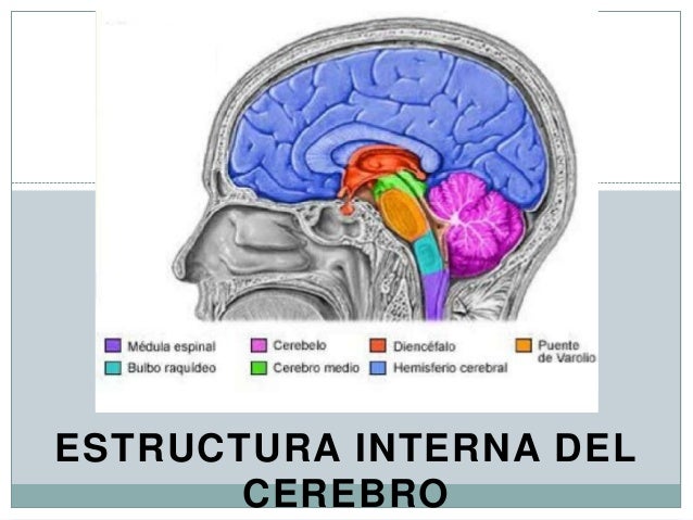
The human brain consists of the cerebrum, the brainstem and the cerebellum. The brain is the organ that controls most of the activities of the body.The brain processes, integrates, and coordinates all of the information it receives from the sense organs.

Imagen relacionadaThe Cerebrum: The cerebrum is the largest part of the human brain, associated with higher brain function such as thought and action.

The Cerebellum: The cerebellum is similar to the cerebrum. This structure is associated with regulation and coordination of movement, posture, and balance.

The brainstem: is the posterior part of the brain, adjoining and structurally continuous with the spinal cord.
The brainstem regulates vital cardiac and respiratory functions and acts as a vehicle for sensory information


SPINAL CORD
Resultado de imagen de spinal cord function 3 eso
The spinal cord is a long, thin, tubular bundle of nervous tissue and support cells that extends from the medulla oblongata in the brainstem to the lumbar region of the vertebral column. The brain and spinal cord together make up the central nervous system (CNS)
The spinal cord has three major functions: as a conduit for motor information, which travels down the spinal cord, as a conduit for sensory information in the reverse direction, and finally as a center for coordinating certain reflexes.


PERIPHERAL NERVOUS SYSTEM

The peripheral nervous system consists of the nerves and nerve ganglia.The main function of the PNS is to connect the CNS to the limbs and organs, essentially serving as a relay between the brain and spinal cord and the rest of the body.The peripheral nervous system is divided into the somatic nervous system, and the autonomic nervous system. The somatic nervous system is under voluntary control, and transmits signals from the brain to end organs such as muscles. The sensory nervous system is part of the somatic nervous system and transmits signals from senses such as taste and touch 


Resultado de imagen de NERVES
NERVES:a group of axons isolate by their myelin sheat and covered in a layer conective tissue.A nerve provides a common pathway for the electrochemical nerve impulses that are transmitted along each of the axons to peripheral organs.
NERVE GANGLION:a cluster nerve cell bodies.Nerve ganglia are 
distributed along the nerve pathway. Ganglia house the cell bodies of afferent nerves and efferent nerves.


NEURON
Is an electrically excitable cell that processes and transmits information through electrical and chemical signals. These signals between neurons occur via specialized connections called synapses. Neurons can connect to each other to form neural networks. Neurons are major components of the brain and spinal cord of the central nervous system.

Resultado de imagen de neuronsDENTRITES are short cells protections with many branches.most neurons nave multiple dentrites.

The AXON is a long cell projection.It is only branched at one end.There is usually only one axon.

The MYELIN SHEAT is a protective covering that surrounds fibres called axons.This sheath is composed of protein and lipids.

The CELL BODY integrates synaptic input and determines the message to be transmitted to other cells by the axon. The cell body also is responsible for a variety of complex biochemical processes.

The NUCLEUS of a neuron is separated from the intracellular fluid and other organelle of the cell, containing the genetic information that guides cellular function.