miércoles, 5 de abril de 2017

DIABETIC NEOPHROPATHY

Nephropathy means kidney disease or damage. Diabetic nephropathy is damage to your kidneys caused by diabetes. In severe cases it can lead to kidney failure.

What causes diabetic nephropathy?

The kidneys have many tiny blood vessels that filter waste from your blood. High blood sugar from diabetes can destroy these blood vessels. Over time, the kidney isn't able to do its job as well. Later it may stop working completely. This is called kidney failure.
Certain things make you more likely to get diabetic nephropathy. If you also have high blood pressure or high cholesterol, or if you smoke, your risk is higher. Also, Native Americans, African Americans, and Hispanics (especially Mexican Americans) have a higher risk.
Resultado de imagen de diabetic nephropathy pdf

What are the symptoms?

There are no symptoms in the early stages. So it's important to have regular urine tests to find kidney damage early. Sometimes early kidney damage can be reversed.
As your kidneys are less able to do their job, you may notice swelling in your body, most often in your feet and legs.

How is it treated?

The main treatment is medicine to lower your blood pressure and prevent or slow the damage to your kidneys. These medicines include:
  • Angiotensin-converting enzyme inhibitors, also called ACE inhibitors.
  • Angiotensin II receptor blockers, also called ARBs.
As damage to the kidneys gets worse, your blood pressure rises. Your cholesterol and triglyceride levels rise too. You may need to take more than one medicine to treat these complications.
And there are other steps you can take. For example:
  • Keep your blood sugar levels within your target range. This can help slow the damage to the small blood vessels in the kidneys.
  • Work with your doctor to keep your blood pressure under control. Your doctor will give you a goal for your blood pressure. Your goal will be based on your health and your age. An example of a goal is to keep your blood pressure below 140/90.
  • Keep your heart healthy by eating healthy foods and exercising regularly. Preventing heart disease is important, because people with diabetes are more likely to have heart and blood vessel diseases. And people with kidney disease are at an even higher risk for heart disease.
  • Watch how much protein you eat. Eating too much is hard on your kidneys. If diabetes has affected your kidneys, limiting how much protein you eat may help you preserve kidney function. Talk to your doctor or dietitian about how much protein is best for you.
  • Watch how much salt you eat. Eating less salt helps keep high blood pressure from getting worse.
  • Don't smoke or use other tobacco products.

This are two foldables about the excretory and urinary system to improve your knowledge in that part of the first floor:


If you want the instructions send me and e-mail almacanosaagenjo@gmail.com

sábado, 1 de abril de 2017


EXCRETORY SYSTEM 

The excretory system is a passive biological system that removes excess, unnecessary materials from the body fluids of an organism, so as to help maintain internal chemical homeostasis and prevent damage to the body. The dual function of excretory systems is the elimination of the waste products of metabolism and to drain the body of used up and broken down components in a liquid and gaseous state. In humans and other amniotes (mammalsbirds and reptiles) most of these substances leave the body as urine and to some degree exhalation, mammals also expel them through sweating.

 PARTS
KIDNEYS:
These are two bean-shaped organs about15 cm long.There is one on either side of spinal column.They are surrounded by a thik layer of  fat.
The kidneys are formed by strucutures called nephrons,wich produce urine.
There is an adrenal gland on top of each kidney.These glands are part of the endocrine system.
Resultado de imagen de excretory systemThe renal artery enters the kidney on its concave side.The renal vein and ureter also leave on this side.

URETERS:
These are two 25 cm-long,narrow tubes.They take urine from this kidneys to the urinary bladder.

URINARY BLADDER:
This is an expandable,muscular organ.Up to half a litre of urine can collect here.
Urine flows continuously from the ureters into the bladder.
When the bladder is full,urination occurs.

URETHRA:Urine flows this tube from the bladder to the outside of the body during urination.


THE NEPHRON

The nephron is the basic structural and functional unit of the kidney.Its functions are vital to life and are regulated by the endocrine system by hormones such as antidiuretic hormonealdosterone, and parathyroid hormone. In humans, a normal kidney contains 800,000 to 1.5 million nephrons.

Resultado de imagen de nephron

FUNCTIONS OF THE NEPHRON
Remove excess water,wastes and other substances from your blood.

Return substances like sodium, potassium or phosphorus whenever any of these substances run low in your body.

Each nephron is composed of two main structures: the glomerulus and renal (kidney) tubule.

The Glomerulus

The glomerulus is a tiny blood vessel or capillary, which looks like a ball of yarn. Actual filtering of your blood occurs in the glomerulus. 

Each of your glomeruli acts like a sieve that helps keep normal proteins and cells in your bloodstream and allows wastes, excess fluid and other substances to pass.


The Tubule

The tubule, also called renal or kidney tubule, is a tiny tube where the wastes, extra fluid and other recyclable substances like sodium and potassium filtered out from the glomerulus pass through.

Your kidneys measure out chemicals like sodium, phosphorus, and potassium and release them back to the blood to return to the body when need arises. In this way, your kidneys regulate the your body’s level of these substances. The right balance is necessary for you to function properly.

THE URINE
Urine is a liquid produced by the kidneys to remove waste products from the bloodstream. Human urine is yellowish in color and variable in chemical composition, but here is a list of its primary components.

Urine samplesPRIMARY COMPONENTS


Human urine consists primarily of water (91% to 96%), with organic solutes including urea, creatinine, uric acid, and trace amounts of enzymes, carbohydrates, hormones, fatty acids, pigments, and mucins, and inorganic ions such as sodium (Na+), potassium (K+), chloride (Cl-), magnesium (Mg2+), calcium (Ca2+), ammonium (NH4+), sulfates (SO42-), and phosphates (e.g., PO43-).

jueves, 9 de marzo de 2017


CIRCULATORY SYSTEM

The circulatory system serves to move blood to a site or sites where it can be oxygenated, and where wastes can be disposed. Circulation then serves to bring newly oxygenated blood to the tissues of the body. As oxygen and other chemicals diffuse out of the blood cells and into the fluid surrounding the cells of the body's tissues, waste produces diffuse into the blood cells to be carried away. Blood circulates through organs such as the liver and kidneys where wastes are removed, and back to the lungs for a fresh dose of oxygen.


 BLOOD

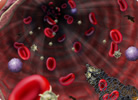
It is the fluid that circulates in the heart, arteries, capillaries, and veins of a vertebrate animal carrying nourishment and oxygen to and bringing away waste products from all parts of the body.
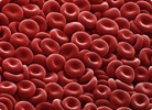
RED BLOOD CELLS
Also called red blood cells or erythrocytes. They are the most numerous cells in the blood. They carry the oxygen from the lungs to the rest of the tissues. The protein that is inside and that binds oxygen is called hemoglobin. Hemoglobin is red and gives this color to the blood.
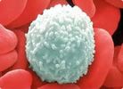


WHITE BLOOD CELLS
Also called leukocytes. They deal with defending the body against the attack of bacteria, viruses, parasites and fungi.


PLATELETS
They are cellular fragments that participate in the protection of the wall of the blood vessels, form a "platelet plug" to prevent bleeding at the site of the injury and produce various substances that help the healing of wounds.


THE PLASMA
It is the liquid part of the blood and is very rich in proteins, among which stand out as the most important: albumin, clotting factors and immunoglobulins.


HEART:
The heart is a pump that circulates blood all around the body.  It is approximately the size of a human fist and is located just to the left of the centre of a human’s chest.

Resultado de imagen de heart biologyThe heart is made of a special type of muscle called the cardiac muscle. Firstly cardiac muscle consists of branching muscle fibres connected to each other in a network, this allows for contractions to begin at one point in the heart and spread outwards in all directions. Secondly cardiac muscle naturally contracts and relaxes rhythmically in “beats”. Thirdly cardiac muscle does not get tired or fatigued even though it is continuously working throughout our lifetime.
The heart is in fact a double pump. The right side of the heart is considered as one pump and the left side of the heart is the second pump. A thick wall called the septum separates the two sides. The right side of the heart carries deoxygenated blood to the lungs to be oxygenated. The left side of the heart pumps oxygenated blood to the rest of the body.

BLOOD VESSELS

Resultado de imagen de arteries
arteries
 
take blood away from the heart to the body organs and tissues. The artery wall is thick and muscular so it can withstand the high pressure of the blood being pumped directly from the heart


capillaries
 are tiny, thin-walled vessels which form a network to take blood through 
the organsand tissues


veins collect blood from the capillaries in the body and return the blood to the heart. The wall of the veins are thin, the blood is at a much lower pressure. To prevent the backflow of this lower pressure blood the veins contain valves

 TYPES OF CIRCULATION

SYSTEMIC CIRCULATION:The blood flows from the left ventricle, through various parts of the body, to the right atrium, i.e. from the left to the right side of the heart through the arteries and veins which traverse the whole body. This circulation is responsible for keeping the body tissues alive by supplying a continuous stream of blood to them.

PULMONARY CIRCULATION:The blood flows from the right ventricle, through the lungs, to the left atrium, i.e. from the right to the left side of the heart. This circulation is responsible for oxygenation of blood. In pulmonary circulation, the blood passes through the lungs where Carbon dioxide is eliminated and Oxygen is added to blood. In this way, the pulmonary circulation makes sure that systemic circulation remains effective.

PORTAL CIRCULATION: It is a part of systemic circulation, which has the following characteristics:
The blood passes through two sets of capillaries before draining into a systemic vein.
The vein draining the first capillary network is known as portal vein which branches like an artery to form the second set of capillaries or sinusoids. Examples: hepatic portal circulation, hypothalamo hypophyseal portal circulation and renal portal circulation.

Tips for having a healthy circulatory system:

-Keep a good intake of protein.

-If you are accustomed to ingesting large amounts of fat in your diet, the most recommended is to decrease it.

-Increases the intake of fruits and vegetables, as they provide the body with the nutrients needed to keep our circulatory system healthy.

-Try to maintain a healthy weight.

-Keep an exercise routine. This will be of great benefit to your general health, for example, to maintain a healthy weight and also for the heart to work and perform the pumping process correctly.

- Avoid maximizing the consumption of alcoholic beverages and also the consumption of cigarettes.

Smoking has been shown to be one of the most prominent reasons for heart attacks.

-Add to your diet omega-3 fatty acids. They will help clear the blood of triglycerides. These can be found in various types of fish such as tuna, verdel, salmon, among others.


miércoles, 8 de marzo de 2017


Today in the hospital, there was a patient that told my that :

Hi! My name is Andrea and I am 25 years old. I´m a soldier and when I was carrying out surveillance work in yhe miuntains, I fell on an embankment, beating my chest with a tree. I have respirartory difficulties, with rapid and superficial breathing, and acute pain that increasese with inspiration. The pain radiates to the shoulder and back and I have a dry, persistent cough with sweating, pallor, hypothension and tachycardia.


PNEUMOTHORAX


Andrea presents respiratory difficultes with rapid and superficial breathing and acute chest pain that increases with inspiration.She also presents sweating,pallor,hypotension andtachycardia.The lungs have membranous coating called pleurae.

These pleurae are of two types:
Resultado de imagen de pneumothorax symptomsThe plarietal pleura and visceral pleura.When air is introduced between both membranes,both pleurae move away ,causing pneumothorax.The lungs are located in the rib cage that form part of the respiratory system.
Next to the lungs you can find the trachea bronchi bronchioles and alveoli.Among the causes of pneumothorax we could find two types:

Intrathoracic.the function of pleura ,wich always producing a certain amount of fluid,is to allow the lung to be properly displaced into de pleural cavity with respiratory movements.The pleural cavity is composed of layers of the membranes lining the lungs and thoracic cavity.


PREVENT:there s no known way to prevent the anset of pneumotorax ;however ,smoking can be avoided in case of patients with this addition.

TREATMENT:the pupose of the tratment is for the lung to expand ,and to reattach the two membranous coating or pleurae.If the pneumothorax is more intense ,it can be treated by evacuating air from the slit by aspiration with a needle.In severe situations,it is recommended to undergo surgery.

martes, 7 de marzo de 2017

LAB

Last day we were doing some lab work at the hospital.I know that I didn´t splain anything about heart and lungs yet but if you want to pratice some exercices you can watch this video and anwser the questions:

video: https://youtu.be/oHMmtqKgs50

video: https://youtu.be/3oYFCQvC-0Q
QUESTIONS:
Explica las principales diferencias entre un pulmón de cordero y uno de humanno:

La estructura es la misma pero el pulmón de el cordeo se adapta anatómicamente a su cuerpo.
¿Qué función tienen los anillos de la tráquea y de los bronquiolos?

Los anillos cartilaginosos constituyen el sostén de la mucosa traqueal e impiden el colapso y los bronquios conducen el aire a los bronquiolos y luego a los alveolos pulmonares.


miércoles, 1 de marzo de 2017

Resultado de imagen de cellular respirationThe processes of cellular respiration and breathing are part of the same process. When you inhale, carbon dioxide is replaced by oxygen in your blood; this oxygen is required by cells to perform aerobic cellular respiration as opposed to anaerobic cellular respiration--a similar process that does not require oxygen.


Breathing

When you inhale, air is drawn into your lungs by the movement of your diaphragm and other breathing muscles. Air enters your lungs via your nose or mouth and down your windpipe to be divided into each lung by your trachea. As air enters the lung, it is channeled down ever smaller pathways, similar in shape to an upside-down tree. At the end of these pathways are tiny air sacs called alveoli. It is here that oxygen in your inhaled air is transferred into your blood while carbon dioxide, a byproduct of your cells breaking down nutrients, is excreted, replacing the oxygen. As you exhale, this carbon dioxide is expelled into the environment. This process is repeated in every breath you take.
CELLULAR RESPIRATION
Oxygen drawn into your blood by your lungs is used in the process of aerobic cellular respiration, an "exergonic" process meaning it produces energy for your body. This energy is drawn from glucose in your cells and is released as a substance known as adenosine triphosphate (ATP). ATP is a form of energy that cells can use to perform their basic functions. In simple terms: glucose + oxygen = carbon dioxide, water and ATP (energy).