jueves, 9 de marzo de 2017


CIRCULATORY SYSTEM

The circulatory system serves to move blood to a site or sites where it can be oxygenated, and where wastes can be disposed. Circulation then serves to bring newly oxygenated blood to the tissues of the body. As oxygen and other chemicals diffuse out of the blood cells and into the fluid surrounding the cells of the body's tissues, waste produces diffuse into the blood cells to be carried away. Blood circulates through organs such as the liver and kidneys where wastes are removed, and back to the lungs for a fresh dose of oxygen.


 BLOOD

It is the fluid that circulates in the heart, arteries, capillaries, and veins of a vertebrate animal carrying nourishment and oxygen to and bringing away waste products from all parts of the body.
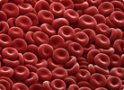
RED BLOOD CELLS
Also called red blood cells or erythrocytes. They are the most numerous cells in the blood. They carry the oxygen from the lungs to the rest of the tissues. The protein that is inside and that binds oxygen is called hemoglobin. Hemoglobin is red and gives this color to the blood.
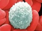


WHITE BLOOD CELLS
Also called leukocytes. They deal with defending the body against the attack of bacteria, viruses, parasites and fungi.


PLATELETS
They are cellular fragments that participate in the protection of the wall of the blood vessels, form a "platelet plug" to prevent bleeding at the site of the injury and produce various substances that help the healing of wounds.
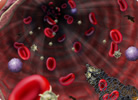

THE PLASMA
It is the liquid part of the blood and is very rich in proteins, among which stand out as the most important: albumin, clotting factors and immunoglobulins.


HEART:
The heart is a pump that circulates blood all around the body.  It is approximately the size of a human fist and is located just to the left of the centre of a human’s chest.

Resultado de imagen de heart biologyThe heart is made of a special type of muscle called the cardiac muscle. Firstly cardiac muscle consists of branching muscle fibres connected to each other in a network, this allows for contractions to begin at one point in the heart and spread outwards in all directions. Secondly cardiac muscle naturally contracts and relaxes rhythmically in “beats”. Thirdly cardiac muscle does not get tired or fatigued even though it is continuously working throughout our lifetime.
The heart is in fact a double pump. The right side of the heart is considered as one pump and the left side of the heart is the second pump. A thick wall called the septum separates the two sides. The right side of the heart carries deoxygenated blood to the lungs to be oxygenated. The left side of the heart pumps oxygenated blood to the rest of the body.

BLOOD VESSELS

Resultado de imagen de arteries
arteries
 
take blood away from the heart to the body organs and tissues. The artery wall is thick and muscular so it can withstand the high pressure of the blood being pumped directly from the heart


capillaries
 are tiny, thin-walled vessels which form a network to take blood through 
the organsand tissues


veins collect blood from the capillaries in the body and return the blood to the heart. The wall of the veins are thin, the blood is at a much lower pressure. To prevent the backflow of this lower pressure blood the veins contain valves

 TYPES OF CIRCULATION

SYSTEMIC CIRCULATION:The blood flows from the left ventricle, through various parts of the body, to the right atrium, i.e. from the left to the right side of the heart through the arteries and veins which traverse the whole body. This circulation is responsible for keeping the body tissues alive by supplying a continuous stream of blood to them.

PULMONARY CIRCULATION:The blood flows from the right ventricle, through the lungs, to the left atrium, i.e. from the right to the left side of the heart. This circulation is responsible for oxygenation of blood. In pulmonary circulation, the blood passes through the lungs where Carbon dioxide is eliminated and Oxygen is added to blood. In this way, the pulmonary circulation makes sure that systemic circulation remains effective.

PORTAL CIRCULATION: It is a part of systemic circulation, which has the following characteristics:
The blood passes through two sets of capillaries before draining into a systemic vein.
The vein draining the first capillary network is known as portal vein which branches like an artery to form the second set of capillaries or sinusoids. Examples: hepatic portal circulation, hypothalamo hypophyseal portal circulation and renal portal circulation.

Tips for having a healthy circulatory system:

-Keep a good intake of protein.

-If you are accustomed to ingesting large amounts of fat in your diet, the most recommended is to decrease it.

-Increases the intake of fruits and vegetables, as they provide the body with the nutrients needed to keep our circulatory system healthy.

-Try to maintain a healthy weight.

-Keep an exercise routine. This will be of great benefit to your general health, for example, to maintain a healthy weight and also for the heart to work and perform the pumping process correctly.

- Avoid maximizing the consumption of alcoholic beverages and also the consumption of cigarettes.

Smoking has been shown to be one of the most prominent reasons for heart attacks.

-Add to your diet omega-3 fatty acids. They will help clear the blood of triglycerides. These can be found in various types of fish such as tuna, verdel, salmon, among others.


miércoles, 8 de marzo de 2017


Today in the hospital, there was a patient that told my that :

Hi! My name is Andrea and I am 25 years old. I´m a soldier and when I was carrying out surveillance work in yhe miuntains, I fell on an embankment, beating my chest with a tree. I have respirartory difficulties, with rapid and superficial breathing, and acute pain that increasese with inspiration. The pain radiates to the shoulder and back and I have a dry, persistent cough with sweating, pallor, hypothension and tachycardia.


PNEUMOTHORAX


Andrea presents respiratory difficultes with rapid and superficial breathing and acute chest pain that increases with inspiration.She also presents sweating,pallor,hypotension andtachycardia.The lungs have membranous coating called pleurae.

These pleurae are of two types:
Resultado de imagen de pneumothorax symptomsThe plarietal pleura and visceral pleura.When air is introduced between both membranes,both pleurae move away ,causing pneumothorax.The lungs are located in the rib cage that form part of the respiratory system.
Next to the lungs you can find the trachea bronchi bronchioles and alveoli.Among the causes of pneumothorax we could find two types:

Intrathoracic.the function of pleura ,wich always producing a certain amount of fluid,is to allow the lung to be properly displaced into de pleural cavity with respiratory movements.The pleural cavity is composed of layers of the membranes lining the lungs and thoracic cavity.


PREVENT:there s no known way to prevent the anset of pneumotorax ;however ,smoking can be avoided in case of patients with this addition.

TREATMENT:the pupose of the tratment is for the lung to expand ,and to reattach the two membranous coating or pleurae.If the pneumothorax is more intense ,it can be treated by evacuating air from the slit by aspiration with a needle.In severe situations,it is recommended to undergo surgery.

martes, 7 de marzo de 2017

LAB

Last day we were doing some lab work at the hospital.I know that I didn´t splain anything about heart and lungs yet but if you want to pratice some exercices you can watch this video and anwser the questions:

video: https://youtu.be/oHMmtqKgs50

video: https://youtu.be/3oYFCQvC-0Q
QUESTIONS:
Explica las principales diferencias entre un pulmón de cordero y uno de humanno:

La estructura es la misma pero el pulmón de el cordeo se adapta anatómicamente a su cuerpo.
¿Qué función tienen los anillos de la tráquea y de los bronquiolos?

Los anillos cartilaginosos constituyen el sostén de la mucosa traqueal e impiden el colapso y los bronquios conducen el aire a los bronquiolos y luego a los alveolos pulmonares.


miércoles, 1 de marzo de 2017

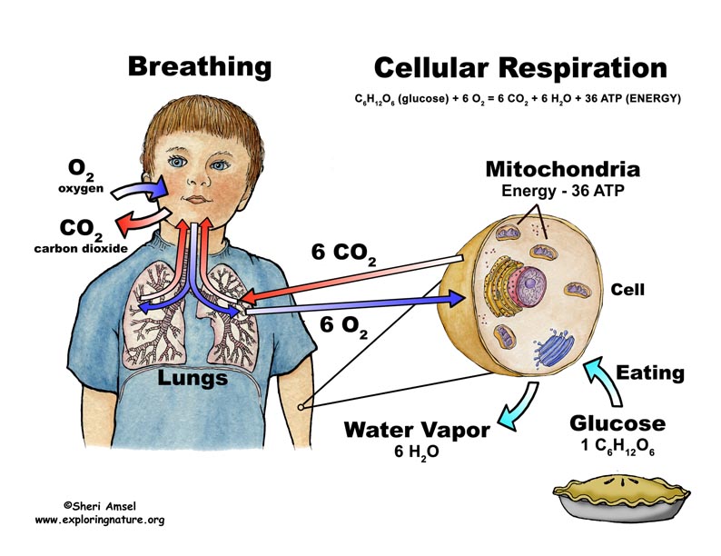
Resultado de imagen de cellular respirationThe processes of cellular respiration and breathing are part of the same process. When you inhale, carbon dioxide is replaced by oxygen in your blood; this oxygen is required by cells to perform aerobic cellular respiration as opposed to anaerobic cellular respiration--a similar process that does not require oxygen.


Breathing

When you inhale, air is drawn into your lungs by the movement of your diaphragm and other breathing muscles. Air enters your lungs via your nose or mouth and down your windpipe to be divided into each lung by your trachea. As air enters the lung, it is channeled down ever smaller pathways, similar in shape to an upside-down tree. At the end of these pathways are tiny air sacs called alveoli. It is here that oxygen in your inhaled air is transferred into your blood while carbon dioxide, a byproduct of your cells breaking down nutrients, is excreted, replacing the oxygen. As you exhale, this carbon dioxide is expelled into the environment. This process is repeated in every breath you take.
CELLULAR RESPIRATION
Oxygen drawn into your blood by your lungs is used in the process of aerobic cellular respiration, an "exergonic" process meaning it produces energy for your body. This energy is drawn from glucose in your cells and is released as a substance known as adenosine triphosphate (ATP). ATP is a form of energy that cells can use to perform their basic functions. In simple terms: glucose + oxygen = carbon dioxide, water and ATP (energy).

domingo, 26 de febrero de 2017

RESPIRATORY SYSTEM
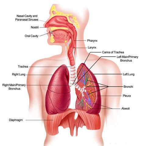
The respiratory system is the manager of providing the oxygen that the body needs and to eliminate the carbon dioxide or carbonic gas that takes place in all the cells across the process called breathing. The breathing is an involuntary and automatic process, in which there is extracted the oxygen of the inspired air and the gases of waste are expelled by the exhaled air. The organs that form a part of the respiratory system are: nose, pharynx, larynx, trachea, bronchi, lungs and diaphragm.

    In the process of respiration the following organs participate:

NOSE :It consists of two large cavities whose function is to allow the entry of air, which is moistened, filtered and heated to a certain temperature through structures called turbinates.

PHARINX:A muscular, membranous duct that helps air to flow into the lower airways.

Resultado de imagen de respiratory systemEPIGLOTTIS :Lid that prevents food from entering the larynx and trachea when swallowing. It also marks the boundary between the oropharynx and the laryngopharynx.

LARINX :Duct whose main function is the filtration of inspired air. In addition, it allows the passage of air to the trachea and lungs and closes to not allow the passage of food during swallowing if the own has not desired and has the function of a vocal organ, ie produces the sound.

WINDPIPE :It provides an open pathway to the air inhaled and exhaled from the lungs.

BRONCHUS :It conducts air from the trachea to the bronchioles.

BRONCHIOLE :It conducts the air that goes from the bronchi through the bronchioles and ending in the alveoli.

SOCKET :Hematosis (Allows gas exchange, that is, inside the blood eliminates carbon dioxide and collects oxygen).

LUNGS :The function of the lungs is to perform the gas exchange with the blood, so the alveoli are in close contact with capillaries.

INTERCOSTAL MUSCLES :The main function of the respiratory muscles is to mobilize a volume of air that serves, after an appropriate gas exchange, to supply oxygen to different tissues.

DIAPHRAGM :Striated muscle separating the thoracic cavity from the abdominal cavity. Intervenes in breathing, lowering the pressure inside the thoracic cavity and increasing the volume during inhalation and increasing the pressure and decreasing the volume during exhalation. This process is carried out, mainly, by the contraction and relaxation of the diaphragm.

miércoles, 22 de febrero de 2017

few days ago in the first floor it was a patient that said that:Hi doctor! I'm Lola Hernandez, I'm 36 years old  and I am slightly overweight . Last weekend, after a family meal, I experienced gradual abdominal pain in the right upper quadrant with irradiation to the back. After that I started to vomit and it become more intense so I went to the hospital, Floridablanca Hospital,where Murphy was detected positive. There was no fever and abdominal ultrasound revealed a distented gallbladder with thickening and hyperemia of its wall, lithiasis and dilated biliary tract. I really don't understand what I have so I need your help. I hope you can help me.

CHOLELITHIASIS
My pacient has cholelithiasis that involves the presence of gallstones, which are concretions that form in the biliary tract, usually in the gallbladder. The gallbladder is a pear-shaped, hollow structure located under the liver and on the right side of the abdomen. Its primary function is to store and concentrate bile, a yellow-brown digestive enzyme produced by the liver and it is part of the biliary tract.


-The thing that causes the illness is her weight, she is overweight and this is one the biggest risk factors, overweight can raise the cholesterol level and also make it harder for the gallbladder to empty completely.


- The common symptoms of cholelithiasis are:

Adominal pain (typically localized in the upper quadrant of the abdomen).
Abdominal swelling, distention or bloating.
Abdominal tenderness.
Clay-colored stools.
Fever and chills.
Loss of appetite.
Nausea with or wihout vomiting.
Pain that radiates from the abdomen to the right shoulder or back.
Yellowing of the skin and whites of the eyes (jaundice)
Abdominal swelling,distention or bloating.
High fever (higher than 101 degrees Fahrenteir)
Severe abdominal pain.


- Lola condition is very serious because she is overweight, she experienced gradual abdominal pain in the right upper quadrant with irradiation to the back, the pain continued with vomiting and became more intense so she went to the hospital where distented gallbladder was revealed  with thickening and hyperemia of its wall and dilated biliary tract. She does not has fever.


Resultado de imagen de CHOLELITHIASIS

- If you have recurrent episodes of cholelithiasis, the most common treatment is surgery to remove the gallbladder. There are two methods: open cholecystectomy, the whole organ is removed through a single incision and laparoscopic technique , when the gallbladder is removed in pieces through several smaller incisions. The second one is the most used  because the gallbladder is not an essential organ.
Other treatments is: Medication to disolve gallstones, it takes a long time, and gallstones may recur.


- If the pacients who have cholelithiasis don't follow the treatment serious complications if gallstones lodge in the common bile duct, which is the large duct formed where the major gallbladder bile duct.


FOLDABLE DIGESTIVE SYSTEM


That is a foldable  that you can do to improve in the standar of digestive system.I´ve you want to do it you only have to send my and email  almacanosaagenjo@gmail.com and i´ll give you all that you need to do it.If you find it a bit difficult you can read my last blog called digestive system in wich I explain all you need to do it.
Here are some photos of what does it look when you finish it:


And if you´ve got any cuestions please talk with me.
                               KISSES 💕💕